In a high-throughput emergency block, a standard posterior-anterior (PA) chest X-ray is often the initial diagnostic line of defense. However, when clinical volume outpaces specialist staff, non-radiologist clinicians must make rapid triage decisions.
Computer-Aided Detection (CAD) software leveraging convolutional neural networks (CNNs) has emerged not to replace the radiologist, but to act as a diagnostic safety net against critical interpretive errors.
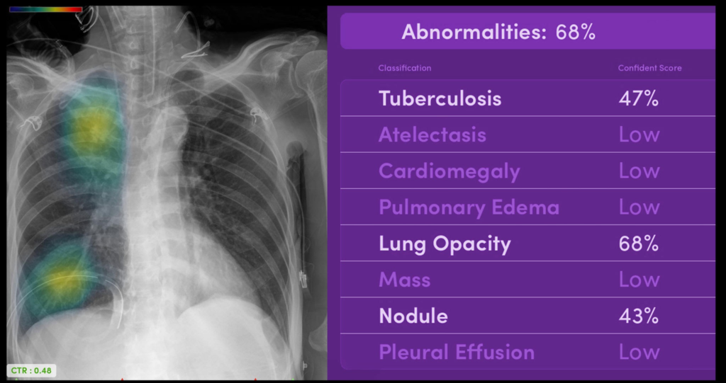
1. The Core Technology: Pixel-Level Visual Attention
Modern radiology AI tools pass raw DICOM imaging data through deep neural networks trained on millions of curated radiograph pairs.
Instead of presenting a binary “normal vs. abnormal” diagnostic result, the system uses two distinct analytical mechanisms:
- Heatmap Localization: Utilizing Grad-CAM (Gradient-weighted Class Activation Mapping) frameworks to project visual heatmaps directly over areas of high diagnostic suspicion (e.g., subtle apical pneumothorax lines or hidden retrocardiac opacities).
- Bone Suppression Algorithms: Temporarily generating a software-modified image that digitally suppresses clavicular and rib shadows, allowing clinicians to look past osseous structures for underlying parenchymal nodules.
2. Clinical Trial Metrics & Diagnostic Safety
When auditing CAD systems against gold-standard consensus panels, the software demonstrates substantial utility in minimizing observational oversight:
| Diagnostic Target | Sensitivity | Specificity | Clinical Value Field |
| Pneumothorax | 94.2% | 91.5% | Critical for trauma and post-procedural triage. |
| Pulmonary Nodules | 88.7% | 86.1% | Catching incidental early-stage malignancies. |
| Pleural Effusion | 92.1% | 93.4% | Accelerating bedside drainage decisions. |
3. The Pitfall: Automation Bias and Confounding Artifacts
Despite exceptional raw metrics, frontline implementation reveals a distinct psychological hurdle: automation bias.
When working long consecutive shifts, overworked residents are susceptible to over-relying on algorithmic flags. It is vital to note that external medical hardware artifacts—such as overlying ECG leads, central venous catheters, or external dress buttons—can cause pixel anomalies that trigger false positive heatmaps.
4. Neural Rounds Recommendation Checklist
For departments seeking to deploy a CXR CAD tool into their triaging interface, our clinical informatics review stresses three deployment parameters:
- Zero-Click Workflow integration: The AI evaluation must run in the background via PACS (Picture Archiving and Communication System) and push notifications straight to the emergency terminal within 60 seconds of image acquisition.
- Explicit Probability Sliders: The interface must allow clinicians to adjust threshold settings to scale down false-positive noise in low-risk cohorts.